
Appearance
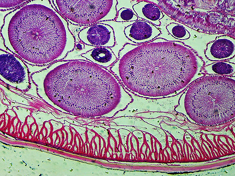
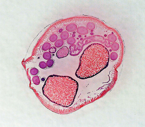
''Ascaris lumbricoides'' is characterized by its great size. Males are 2–4 mm in diameter and 15–31 cm long. The male's posterior end is curved ventrally and has a bluntly pointed tail. Females are 3–6 mm wide and 20–49 cm long.The vulva is located in the anterior end and accounts for about one-third of its body length. Uteri may contain up to 27 million eggs at a time, with 200,000 being laid per day. Fertilized eggs are oval to round in shape and are 45–75 μm long and 35–50 μm wide with a thick outer shell. Unfertilized eggs measure 88–94 μm long and 44 μm wide.
Evolution
Giant intestinal roundworms have been known since antiquity. In 1758 Linnaeus named them ''Ascaris lumbricoides''. For many centuries, they were thought to arise by spontaneous generation. In 1855, ''Ascaris'' eggs were found in human faeces by Henry Ransom in England then this was described in the literature two years later by Casimir-Joseph Davaine in France. Attempts to infect animals by feeding them eggs were unsuccessful. In 1886, Salvatore Calandruccio in Italy successfully infected a boy to whom he had given 150 eggs. Battista Grassi published this information without giving any acknowledgement to Calandruccio. Development was thought to occur directly within the bowel lumen but in Francis Stewart in Hong Kong in 1916 fed eggs to rats, then later mice, and found infective larvae in the faeces and in the lungs but no mature worms. In 1918, Sadao Yoshida ingested larvae recovered from the trachea of a guinea pig, then found eggs in his own stools 76 days later. In 1922, Shimesu Koino ingested 2,000 ''Ascaris lumbricoides'' eggs, found larvae in his sputum a few days later, then after 50 days took an anthelmintic and recovered 667 immature ''Ascaris lumbricoides'', thus confirming the life cycle.References:
Some text fragments are auto parsed from Wikipedia.